Using patient-specific 3D or virtual reality (VR) models for preoperative assessment could help anesthesiologists better plan for procedures involving patients with suspected difficult airways, according to an abstract presented at the Euroanaesthesia 2021 meeting in Munich, Germany.
“With a specific plan in place, there is less trial and error, wastage of equipment and trauma to the patient,” the authors write.
Although these modeling technologies have been used to train residents in intubation procedures, they are not as widely used in clinical anesthesia, said lead author Ruth Shaylor, BMBS, an anesthesiologist at the Tel Aviv Medical Center in Israel. “Until now, we hadn’t realized how it can be useful for us,” she told Medscape Medical News.

Dr Ruth Shaylor
That all changed when a new thoracic surgeon joined the hospital and brought with him pediatric patients, a group with whom Shaylor and colleagues had not previously worked. “Suddenly, we were presented with a patient where in theory, we knew what to do, but in practice, we’d never done it before,” she said. Because she already regularly used 3D-printing to train residents, she made the next logical leap: “If I print off tracheas to teach the residents how to intubate, why don’t I print a patient-specific trachea to teach myself?” she recalled.
Using medical imaging from one patient, she printed a 3D-model of the patient’s trachea that allowed her to select the correct equipment and practice the procedure. Soon after, she used 3D-printing again to help plan the intubation procedure for an adult patient with severe narrowing of the trachea. “After those couple of cases, the entire service took off,” Shaylor noted.
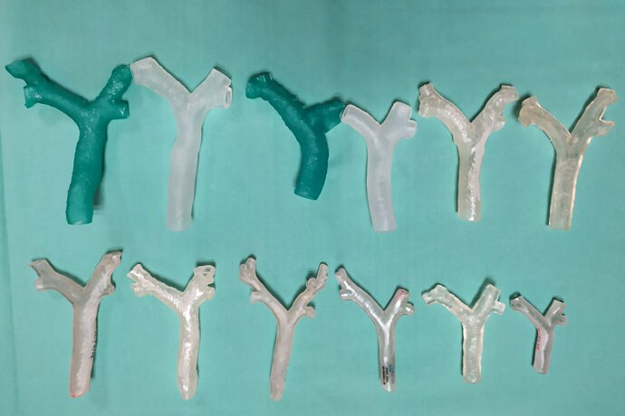
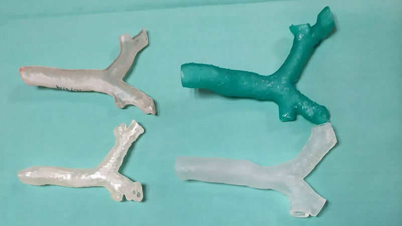
For 3D printed airway models, the team from Tel Aviv Medical Center in Israel started with a CT of the trachea, main bronchi, and lobar bronchi.
The department now uses 3D-modeling for pediatric patients needing thoracic surgery, patients with difficult airways, or patients with lung or mediastinal masses. To assess the potential benefits of the program, Shaylor and colleagues conducted a review of all cases referred for 3D-modeling during the first 2 years of the program, from July 2019 to July 2021. Over the 2-year period, 10 pediatric, two fetal, and eight adult cases were referred to the program. Seventeen patients presented with suspected difficult airways and three had anterior mediastinal masses.
For 3D printed airway models, the team started with a CT of the trachea, main bronchi, and lobar bronchi. These images were then imported into the post-processing software, and the 3D model was then printed — building outward to maintain the internal diameter of the model — in clear plastic. The treating anesthesiologist would then use the model to determine the most appropriate airway plan. The virtual reality models were constructed from volume-rendered medical images — either CT or MRI — and viewed on Oculus VR headsets. The cardiothoracic surgeons responsible for extracorporeal membrane oxygenation (ECMO) and treating anesthesiologists jointly reviewed the VR model and the induction and anesthesia plans were devised together.
The team printed 15 3D models and created five VR reconstructions over the study period. Thirteen of the 15 cases using 3D printed models used the plan devised in the model as the final airway plan. The additional two cases had poor initial imaging, and the devised plan on the model was more conservative than the final airway plan. After reviewing the VR images, none of the three patients presenting with mediastinal masses had preinduction ECMO or wires inserted.

Dr Ian Chao
“Although a small observational study with only 20 cases, it is the first attempt I have seen of a center actively formalizing a pathway for ‘routine use’ of this technology to aid clinical decision-making,” said Ian Chao, MBBS, a specialist anesthetist in Melbourne, Australia, in an email interview with Medscape. His research focuses on 3D-printing in anesthesia, and he was not involved with the work. “Although we are still in very early days for research in this field, hopefully this abstract helps to continue to build traction and encourages other clinicians to consider this exciting technology as viable options to explore for anesthesiology.”
Seth Friedman, PhD, the manager of Innovation Imaging and Simulation Modeling at Seattle Children’s in Washington, who was also unaffiliated with the work, agreed with Chao, noting that the abstract suggests that communication — both between providers and between the provider and patient — improved with the use of 3D modeling. The next step, he said, is measuring outcomes in procedures that used 3D modeling tools vs those that did not.
Although the authors indicate that the models improved care by decreasing trial and error as well as trauma to the patient, more data is necessary to back up these statements, Friedman said. “If for every complex airway patient you made a 3D-printed or VR model, and it results in consistently less complications and better outcomes” that could help spur greater investment in these modeling technologies in anesthesia care, “that’s the data that we need to generate to move this forward as a field.”
Friedman has been part of the beta testing program for Stratasys’ new digital anatomy creator software. This software was not used in the research, but the team did utilize a Stratasys 3D printer. Shaylor and Chao have disclosed no relevant financial relationships.
Euroanaesthesia 2021. Abstract: Virtual reality and 3D printing in clinical anesthesia; 18 months experience in a large, single medical center. Presented December 18, 2021.
For more news, follow Medscape on Facebook, Twitter, Instagram, YouTube, and LinkedIn