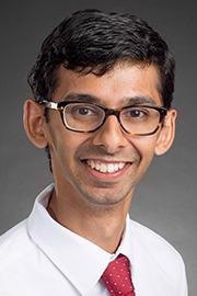
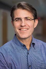
 Thought LeadersAnand Patel and Michael Dyer Pediatric Oncologist and Developmental BiologistSt. Jude Children’s Hospital
In this interview, News-Medical speaks to Anand Patel and Michael Dyer from St. Jude’s Children’s Research Hospital about their recent research suggesting EGFR inhibitors may prevent rhabdomyosarcoma recurrence.
Please can you introduce yourself, tell us about your scientific background, and what inspired your latest research?
Anand Patel: I am a pediatric oncologist at St. Jude Children’s Hospital. I attended college in the Pratt School of Engineering at Duke University and earned MD and Ph.D. doctorates from the Mayo Clinic. I then completed a pediatric residency at St. Louis Children’s Hospital, followed by a Pediatric Hematology-Oncology fellowship at St. Jude.
This work was inspired by questions that arose when I took care of patients. As a pediatric oncologist, I specialize in caring for children with sarcomas, which are tumors of the bone and soft tissues. Often, my patients had a terrific response to treatment; but unfortunately, months or years later, their tumors came back. To me, that meant a tiny fraction of the cancer cells are able to survive therapy, and I wanted to understand what those surviving cells are. So, I joined Michael Dyer’s lab to see if we could build the tools to map the cells within rhabdomyosarcoma and figure out which cells survive therapy. The hope is that, by understanding this process, we can find more effective treatments that completely wipe out tumors without leaving those rare cancer cells behind.
Mike Dyer: I am a developmental biologist interested in understanding how developmental pathways are hijacked in pediatric cancer. I earned my bachelor’s degree at UCLA, then did my Ph.D. at Harvard University and postdoctoral training at Harvard Medical School. While I am a basic researcher, my lab also performs translational research to advance the most promising novel therapeutics to the clinic.
The inspiration for the most recent work came 10 years ago when we performed genome sequencing of rhabdomyosarcoma. We learned that there was profound clonal selection with treatment but didn’t understand anything about the cells that survived chemotherapy. That led to a decade-long journey to identify those cells that survive treatment and contribute to disease recurrence. We are now applying this approach to all pediatric solid tumors to be more effective at preventing recurrence and improving outcomes.
Rhabdomyosarcoma is a rare type of soft tissue cancer but the most common type of soft tissue sarcoma in children. Could you tell us a bit more about the pathology of rhabdomyosarcoma?
Anand: Rhabdomyosarcoma is the most common soft tissue sarcoma in childhood, and about 400 children in the United States get newly diagnosed with this disease every year. The disease gets its name from the fact that these tumors look like immature muscle under the microscope. These tumors can form almost anywhere in the body, including around the eye, neck, abdomen, or limbs. In children, we see two major subtypes of rhabdomyosarcoma: (1) the embryonal variant, often called the “fusion-negative” subtype, and (2) the alveolar variant, often called the “fusion-positive” subtype. The fusion-positive subtype has a unique genomic alteration where two transcription factors (PAX3 or PAX7 on one side and FOXO1 on the other side) are fused.
Mike: It has always been interesting that the alveolar type of rhabdomyosarcoma represents a later stage(s) of muscle development. In this study, we were able to extend that concept to single tumor cells and show that the most immature mesoderm-like cells are enriched in the embryonal subtype.
Image Credit: Buravleva stock/Shutterstock.com
Rhabdomyosarcoma can reoccur after therapy. What are the current therapies used to treat this soft tissue cancer, and what are the outcomes and prognostic factors after recurrence?
Anand: Treatment for rhabdomyosarcoma has centered around chemotherapy combined with surgery and/or radiation. As you can imagine, these therapies can be toxic; even for those kids that get cured, they live with lifelong effects from treatment. To make matters worse, children who have intermediate- or high-risk rhabdomyosarcoma, whose tumor can’t be surgically removed or whose tumors have already spread, have very poor outcomes (5-year overall survival less than 30%). Disappointingly, we as a community have struggled to find safe and effective treatments that improve the survival rates for these patients.
Mike: Looking ahead, we are mobilizing all options to achieve our concept of total clonal therapy. We need to eliminate all the tumor cells (each clone) to achieve a cure. We will employ chemotherapy using targeted agents (e.g., EGFR inhibitors) and antibody-drug conjugates and even consider CAR T-cells. This may be an ideal situation for immune-oncology because the number of cells that remain after treatment is relatively small, so using the immune system to kill the remaining cells is an important strategy moving forward.
You studied a population of cells that persists after therapy, causing rhabdomyosarcoma recurrence. How did you undertake this research, and what were your main findings?
Anand: We took advantage of new single-cell sequencing technologies, which have revolutionized cancer biology over the last five years. These tools allow us to profile cells within a tumor individually and give us an unprecedented amount of information about how tumors are structured. Using these techniques, we were able to create an “atlas” of the cancer cells within 18 patient rhabdomyosarcomas. We found three different types of cells that mimic different stages of muscle development, from primordial “mesoderm-like” cells to intermediate “myoblast-like” cells to mature “myocyte-like” cells. We were able to use experimental models generated from those patients to start charting what happens to tumors as we exposed them to chemotherapy.
To our surprise, one of the three cell types, the mesoderm-like cells, was resistant to chemotherapy, and these cells seemed to have EGFR signaling activity. As a proof of principle, we showed that combining chemotherapy with EGFR inhibitors could improve the treatment outcomes in cells grown as organoids (3D clusters of cancer cells) or in mice implanted with patient tumors (xenografts).
Mike: This is one of the most important discoveries in pediatric solid tumor research in the past ten years. There has been little improvement in outcomes for decades, and we have shown that the clinical trials have not improved survival because they were targeting the same cell populations and missing the rare cell population that was evading therapy. Now that we know the molecular and cellular mechanisms of recurrence, we can do a better job of killing all the tumor cells and preventing recurrence.
What advantages did the newly available techniques used in this study, such as single-nucleus RNA-sequencing and epigenetic profiling, provide?
Anand: These new techniques made all the difference. Before, we were forced to grind up tumors and look at them as one “bulk” data point. Unfortunately, the rare cells we were looking for would get lost within that bulk data. Without single-cell sequencing technologies, we wouldn’t have been able to dissect each of the cell types. With that information, we were then able to find a treatment that specifically targeted the mesoderm-like cells.
Mike: As I mentioned above, we knew there was clonal selection over a decade ago from genome sequencing. It took all these years and advanced technologies (single-cell RNA-seq) to finally isolate those tumor cells that survive treatment. It is one of those great examples in biomedical research where persistence pays off when a new technology comes on the scene.

Image Credit: Rachaphak/Shutterstock.com
In your study, you mention collaboration with St. Jude Childhood Solid Tumor Network (CSTN) and the Pediatric Cancer Genome Project. How important is collaboration to increasing our understanding of diseases such as rhabdomyosarcoma?
Anand: The Pediatric Cancer Genome Project (PCGP) and Childhood Solid Tumor Network (CSTN) were foundational resources for this study. PCGP characterized the genetic alterations within a huge number of pediatric cancers. CSTN is an ongoing effort to develop patient-derived models of pediatric solid tumors, and it’s generated over 300 models to date; many of these diseases had no patient-derived models before CSTN started.
Both PCGP and CSTN are entirely “open”: all of the data from PCGP is shared openly for the world to see, and all of the animal models from CSTN are available free of charge to scientists around the world. Both of these efforts are designed to cut through the red tape to disseminate information and tools for scientists to continue to build and learn. That spirit of openness inspired us to make all the atlas data available in an easy-to-use visualizer so that other scientists can also use the data we generated.
Mike: Free and open sharing is part of the St. Jude mission, and we share all of our tumor models with researchers around the world. To date, there have been 644 requests from 261 different researchers at 123 institutions across 17 countries. This is one way that we hope to accelerate discoveries in the field of pediatric cancer to improve outcomes.
The population of cells that cause recurrence depends on epidermal growth factor receptor (EGFR) signaling and is sensitive to EGFR inhibitors. How are EGFR inhibitors already used in cancer treatment, and how do you hope they will change the treatment of rhabdomyosarcoma?
Anand: We were pleased to see EGFR as a potential target within our data because there is a deep and long-standing track record of targeting that receptor. Lung cancer doctors have been using inhibitors of EGFR for over 15 years, and they can be safely administered to patients. So, it seemed natural to test EGFR inhibitors against rhabdomyosarcoma. The results were exciting, but we still have a lot of work to do before we’re ready to test EGFR inhibitors in patients.
Mike: In addition to the EGFR inhibitors, we are also testing antibody-drug conjugates and hope to explore the possibility of developing a CAR T cell for this target. It is a perfect situation for immune-oncology because the number of cells that survive treatment is small and have different cell surface proteins that the immune system may be able to recognize and eliminate.
Your work also relied on computational approaches that used machine learning and systems biology. How have you seen these approaches change cancer research, and how do you foresee them continuing to do so in the future?
Anand: I think these computational tools are game-changers, and they’ve already made a major impact on cancer research. These tools allow us to analyze massive datasets that no single person could ever do by hand. Moreover, they help us find patterns that wouldn’t have been obvious to the human eye. We worked with two amazing computational biologists during this study, Dr. Xin Huang and Dr. Xiang Chen. Without Dr. Huang’s systems biology tools, we wouldn’t have found EGFR as a target in mesoderm-like cells; likewise, Dr. Chen’s machine learning tools allowed us to precisely compare normal fetal tissue to rhabdomyosarcoma to show how similar the two are.
It’s remarkable to see the computational field grow. I think the next major hurdle will be to figure out how to put together many different types of data – things like genomics, clinical information, radiography, and histology – to integrate all this information into useful insight. As an oncologist, I dream of the day when I can use these tools to diagnose my patients rapidly and identify the best treatments for them. I think it’ll be hard work to get there, but it will dramatically impact how we deliver care in the future.
Mike: In my opinion, computational biology will have the greatest impact on biomedical research and human health over the next decade. We are very good at generating large amounts of data at high resolution, and the challenge is making sense of those complex datasets. Computational methods, including machine learning, artificial intelligence, and neural networks, are instrumental in making sense of large, complex multidimensional datasets.
What is next for you and your research?
Anand: We are continuing to study these mesoderm-like cells, and I’m particularly interested in finding the most effective way to kill these cells. In addition, the fusion-positive alveolar variant of rhabdomyosarcoma had very few mesoderm-like cells, so we’re investigating whether these tumors have a different mechanism of resistance. Finally, we’re starting to look at other pediatric solid tumors to see whether we can apply this approach to other diseases.
Mike: We want to move new effective treatments into the clinic and extend this concept of rare cells surviving treatment to other pediatric solid tumors. We have all the data at our fingertips and now need to analyze the data and perform the studies to validate the underlying biology. We envision that this will be the foundation for future studies over the next 3-5 years.
Where can readers find more information?
About Anand Patel, MD, PhD
I am an instructor within the Department of Oncology at St. Jude Children’s Research Hospital. I am a pediatric oncologist who treats children with solid tumors. My r esearch interest, as a member of Michael Dyer’s lab, focuses on understanding how pediatric tumors adapt to survive therapy and spread throughout the body. We use a variety of technologies within the lab, including single-cell sequencing techniques and patient-derived animal models of pediatric cancer. I have been awarded a Damon Runyon-Sohn Pediatric Cancer Fellowship and recently was named an AACR Next Gen Star. I am funded by Alex’s Lemonade Stand, Hyundai Hope on Wheels, and the National Pediatric Cancer Foundations.
About Michael Dyer, PhD
Michael Dyer, Ph.D. was originally recruited to work on retinal development in the Department of Developmental Neurobiology at St. Jude Children’s Research Hospital in 2002. He was aware of the hospital’s reputation for cancer research, but was very impressed with the breadth and depth of research across several different disciplines.  Since arriving at St. Jude, his research has moved into new areas because of the unique collaborative opportunities.
He is currently focusing on trying to understand how normal developmental processes go awry in pediatric solid tumors of the eye, bone, muscle and peripheral nervous system. He is hoping to find vulnerabilities in these devastating childhood cancers that can be exploited with new combinations.
Dr. Dyer received his bachelor’s degree from UCLA, his doctoral degree from Harvard University and his postdoctoral training from Harvard Medical School. He has received numerous awards since joining the, including being named a Pew Scholar, the Cogan Award Recipient and an Investigator of the Howard Hughes Medical Institute. In 2014, Dr. Dyer was named the Richard C. Shadyac Endowed Chair in Pediatric Cancer Research.
