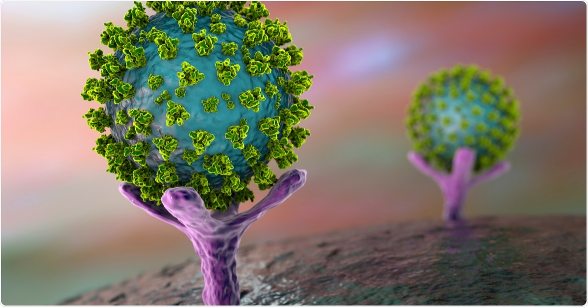
A recent study conducted by a team of international scientists has revealed that severe acute respiratory syndrome coronavirus 2 (SARS-CoV-2), the causative pathogen of coronavirus disease 2019 (COVID-19), suppresses the expression and function of human angiotensin-converting enzyme 2 (ACE2) and induces the expression of interferon-stimulated genes at the initial phase of infection. The study is currently available on the bioRxiv* preprint server.
The entry of SARS-CoV-2 into host cells occurs via ACE2, which is an ectopeptidase and a functional receptor for the spike protein of many human coronaviruses. Since the emergence of the COVID-19 pandemic, many studies have investigated the cellular consequences of spike-ACE2 interaction. Some of these studies have shown that an interferon (IFN)-mediated induction of ACE2 expression occurs upon SARS-CoV-2 infection.
Regarding the mode of action of ACE2, it is known that the ectodomain of ACE2 is cleaved and released from the cell membrane by two proteases, namely ADAM17 and TMPRRS2. Moreover, TMPRRS2 facilitates the entry of SARS-CoV-2 into host cells by priming the spike protein. In the case of SARS-CoV-2 infection, the ectodomain of ACE2, which preserves its catalytic activity even after release from the cell membrane, may act as a soluble decoy to inhibit new viral infections, or may reduce local inflammation through its tissue-protective functions.
To better understand the molecular mechanism of SARS-CoV-2 transmission and pathogenesis, it is important to evaluate the cascade of inflammatory and immune signaling events that occur soon after the induction of SARS-CoV-2 infection.
Current study design
The scientists used nasopharyngeal samples collected from SARS-CoV-2-infected individuals to study the influence of ACE2 on host immune responses. Specifically, they investigated the direct impact of SARS-CoV-2 infection on ACE2 expression and function.
The nasopharyngeal swabs were collected from 40 non-hospitalized COVID-19 patients at 2 points in time: at the time of recruitment (day 0) and 3 days after. As a control, nasopharyngeal samples were also collected from 20 non-infected individuals. A quantitative reverse transcriptase-polymerase chain reaction was used to quantify the viral load, and a fluorescent enzymatic assay was used to analyze the ACE2 activity.
Important observations
The scientists measured the enzymatic activity of soluble ACE2 in nasopharyngeal samples to mimic the ACE2 activity in the upper respiratory tract. They also checked the ACE2 activity in serum samples. The findings revealed that ACE2 activity was significantly lower in nasopharyngeal samples compared to that in serum samples. This indicates potential involvement of ACE2 in the upper respiratory tract.
Another interesting observation was that compared to samples collected at day 0, samples collected on day 3 showed significantly lower ACE2 activity. Moreover, a significantly lower level of ACE2 expression was observed in SARS-CoV-2 positive swab samples. A further reduction in ACE2 expression was observed in day 3 samples. These findings indicate that SARS-CoV-2 is capable of downregulating both the expression and function of ACE2, probably by triggering the cleavage and release of ACE2 from the cell membrane.
Another strong indication of the direct impact of SARS-CoV-2 infection on ACE2 function came from the observation that reduction in ACE2 expression and function was positively correlated with a drop in viral load over time (from day 0 to day 3).
To investigate the mode of action of SARS-CoV-2, the scientists measured the expressions of two main proteases (ADAM17 and TMPRRS2) that catalyze the cleavage of ACE2 ectodomain. They observed a reduction in TMPRRS2 expression in infected samples compared to that in uninfected samples.
While checking the expression on IFN-stimulated genes, they observed significantly higher expressions of DDX58, CXCL10 and IL-6 in SARS-CoV-2-infected samples collected at day 0. However, reduced expressions of these genes were noticed in infected samples collected at day 3. The initial induction of IFN-stimulated gene expression was positively correlated with higher viral load. Taken together, these findings indicate that soon after viral entry, an induction in IFN-mediated antiviral response occurs, which is subsequently suppressed by viral proteins to facilitate survival and infectivity.
Study significance
The findings of the current study contradict previous study findings showing induction of ACE2 expression by IFN upon SARS-CoV-2 infection. Moreover, the study reveals that viral load is associated with the initial induction of antiviral response and that upregulation of IFN-stimulated genes rapidly wanes within a few days of infection induction.
Several studies have shown that the presence of circulating ACE2 at the site of infection may be beneficial in terms of counterbalancing proinflammatory responses. A new role of soluble ACE2 as a blocker of SARS-CoV-2 has recently been identified. Based on the current study findings, the scientists suggest that recombinant ACE2 can be applied locally at the initial phase of infection to control the viral spread.
*Important Notice
bioRxiv publishes preliminary scientific reports that are not peer-reviewed and, therefore, should not be regarded as conclusive, guide clinical practice/health-related behavior, or treated as established information.
