Editor’s note: Find the latest COVID-19 news and guidance in Medscape’s Coronavirus Resource Center.
Background
-
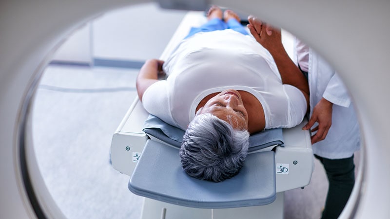
A multicenter study looked into brain hypometabolism detected by PET scans done on patients who had had SARS-CoV-2 infection and were referred for suspected neurologic long COVID.
-
Almost half of the scans were interpreted as abnormal, with more than a quarter of them being interpreted as severely affected.
-
This exam could reassure the half of patients for whom a brain impairment was suspected and enable the other half to get access to adequate care, as well as the requisite social and medical recognition for their condition.
Why Is This Important?
These analyses highlight the fact that brain scans can complement, in routine practice, the examination of patients diagnosed with neurologic long COVID. This proposed pattern is easily applicable for visual interpretation, with consideration required for fronto-basal paramedian regions, the brainstem, and the cerebellum, which are not classic hypometabolic regions reported in other usual disorders (such as neurodegenerative and psychiatric diseases).
Methods
As mentioned above, this was a multicenter study. From August to October 2021, physicians at three French nuclear medicine departments (Nancy Regional University Hospital Center, Nancy, France; Timone Hospital-Marseille Public Hospital System; and Pitié-Salpêtrière Hospital-Paris Public Hospital System) retrospectively reviewed and interpreted brain 18F-FDG PET scans according to the COVID hypometabolic pattern. The scans had been performed on patients who had had SARS-CoV-2 infection and were referred to one of the hospitals for suspected neurological long COVID. The physicians reviewed and classified the scans according to the same visual interpretation analysis.
Main Results
There were a total of 143 patients (mean age, 47.4 years; 98 women). On average, PET scans were performed at 10.9 months from symptom onset. Based on the visual analysis by experienced nuclear physicians, 53% of the scans were interpreted as normal, 21% as mildly to moderately affected, and 26% as severely affected, according to the COVID hypometabolic pattern; the cases had either no involvement, incomplete or mild/moderate involvement, and severe involvement, respectively, of the olfactory bulbs within the fronto-basal region and connected cerebral regions (including other limbic/paralimbic regions, the brainstem, and the cerebellum).
In conclusion, the proposed PET metabolic pattern is easily identified upon visual interpretation in clinical routine for approximately one half of patients with suspected neurologic long COVID, requiring further adapted follow-up and medical care.
This article was translated from French and appeared on Univadis.
For more news, follow Medscape on Facebook, Twitter, Instagram, YouTube, and LinkedIn